
 By
By
- Torsten B. Moeller
- Emil Reif
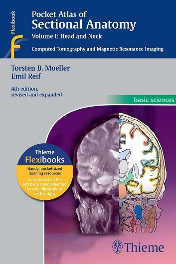
This comprehensive, easy-to-consult pocket atlas is renowned for its superb illustrations and ability to depict sectional anatomy in every plane. Together with Volumes II and III, it provides a highly specialized navigational tool for all clinicians who need to master radiologic anatomy and accurately interpret CT and MR images.
Special features of Pocket Atlas of Sectional Anatomy:
- Didactic organization in two-page units, with high-quality radiographs on one side and brilliant, full-color diagrams on the other
- Hundreds of high-resolution CT and MR images made with the latest generation of scanners (e.g., 3T MRI, 64-slice CT)
- Color-coded schematic drawings that indicate the level of each section
- Concise, easy-to-read labeling of all figures
- Sectional enlargements and magnified views for easy classification of anatomic structures
Updates for the 4th edition of Volume I:
- New cranial CT imaging sequences of the axial and coronal temporal bone
- Expanded MR section, with all new 3T MR images of the temporal lobe and hippocampus, basilar artery, cranial nerves, cavernous sinus, and more
- New arterial MR angiography sequences of the neck and additional larynx images
Compact, easy-to-use, highly visual, and designed for quick recall, this book is ideal for use in both the clinical and classroom settings.
Download
Note: Only Radiology member can download this ebook. Learn more here!
Related Books
 Pocket Atlas of Sectional Anatomy, Volume II: Thorax, Heart, Abdomen and Pelvis Computed Tomography and Magnetic Resonance Imaging, 4th Edition
 Temporal Bone Imaging
 Imaging of the Temporal Bone
 Practical Differential Diagnosis for CT and MRI
 Imaging for Otolaryngologists
 Pocket Atlas of Radiographic Anatomy, 3rd Edition
 Radcases Head and Neck Imaging
 CT of the Head and Spine
 Imaging of the Head and Neck, 2nd Edition
 Atlas of Head and Neck Ultrasound
Thanks for the uploads, can you get Grainger & Allison’s Diagnostic Radiology, 6th Edition
EXPERT CONSULT:
Please
Thanks
Could you please get volume 3?
Thnx
Mediafire download access please!!!
fixed
Link is not working. Thanks!
fixed
Pocket Atlas of Sectional Anatomy
fixed
Invalid link ‘code’.
fixed