By 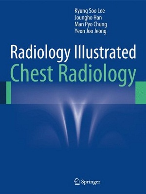
- Kyung Soo Lee (Author),
- Joungho Han (Author),
- Man Pyo Chung (Author),
- Yeon Joo Jeong (Author)
The purpose of this atlas is to illustrate how to achieve reliable diagnoses when confronted by the different abnormalities, or “disease patterns”, that may be visualized on CT scans of the chest. The task of pattern recognition has been greatly facilitated by the advent of multidetector CT (MDCT), and the focus of the book is very much on the role of state-of-the-art MDCT. A wide range of disease patterns and distributions are covered, with emphasis on the typical imaging characteristics of the various focal and diffuse lung diseases. In addition, clinical information relevant to differential diagnosis is provided and the underlying gross and microscopic pathology is depicted, permitting CT–pathology correlation. The entire information relevant to each disease pattern is also tabulated for ease of reference. This book will be an invaluable handy tool that will enable the reader to quickly and easily reach a diagnosis appropriate to the pattern of lung abnormality identified on CT scans.
Download
Note: Only Radiology member can download this ebook. Learn more here!








Plz try for this book friend….
Fundamentals of Fluoroscopy, 1edition
By Houston.
Thank you.
Thank you
great
thanks
great
great works thank u sooooooooo much
Dear admin,
could you find the following title?:
I.C.U. Chest Radiology: Principles and Case Studies
Hardcover: 183 pages
Publisher: Wiley-Blackwell; 1 edition (April 12, 2010)
ISBN-10: 0470450347
ISBN-13: 978-0470450345
Thanks you
http://radiology.downloadmedicalbook.com/4683/i-c-u-chest-radiology-principles-and-case-studies.html
Thnx
link is down sir
fixed