 By
By
- Juergen Kraemer, M.D., Professor, St .-Josef-Hospital, Orthopedic University Clinic, Bochum, Germany
- Odo Koester, M.D., Professor, Department of Radiology, and Nuclear Medicine, St .-Josef-Hospital, Ruhr University, Bochum, Germany
Hundreds of diagnostic images improve your clinical decision-making!
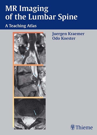
Two-thirds of degenerative diseases of the vertebral column involve the lumbar spine. Magnetic resonance imaging plays a pivotal role in diagnosis and treatment. With more than 450 illustrations and 78 case studies illustrating various constellations of findings, this book provides a wealth of illustrations that guide the reader through the MR imaging of lumbar disk herniations and spinal stenosis:
- Impressive series of MR images illustrate both common and unusual findings, helping to enhance conceptual understanding and sharpen diagnostic perception.
- Clinical findings and progression are covered in addition to MRI findings, helping the reader to appreciate the correlations between clinical and imaging findings.
- The role of diagnostic imaging is addressed for specific disorders, helping to foster the more discriminating use of imaging procedures in the lumbar spine.
The book concludes with a chapter on the current technique of performing CT-guided injections at the lumbar level.
Download
Note: Only Radiology member can download this ebook. Learn more here!


the file is no longer available
???????
the link is not working
fixed
Link dead – can you fix please? Thanks
fixed