 By
By
- Val M. Runge, MD, Robert and Alma Moreton Centennial Chair in Radiology, Department of Radiology, Scott & White Clinic and Hospital, Texas A&M University Health Science Center, Temple, Texas
- Wolfgang R. Nitz, PhD, Senior Patent Manager, Siemens AG, Healthcare Sector, Erlangen, Germany, Department of Radiology, University Hospital of Regensburg, Regensburg, Germany
- Stuart H. Schmeets, BS, RT(R)(MR), 3 T and Cardiac Segment Manager, Siemens Medical Solutions USA, Inc., Malvern, Pennsylvania
Award Winner, RSNA 2009!
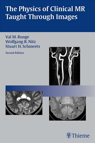
This lavishly illustrated book uses high-quality images to present a practical guide to the physics of magnetic resonance. Written by internationally renowned authors, the book places an emphasis on learning visually through images of real cases rather than through mathematical equations and provides the fundamental information needed to achieve the best images in everyday clinical practice. This edition features new images and incorporates information on the latest technical advances in the field, discussing such important topics as 3 T, specific absorption rate (SAR), arterial spin labeling, continuous moving table MR, and time-resolved contrast enhanced MR angiography.
Highlights:
- Concise chapters make difficult concepts easy to digest
- 400 high-quality images and illustrations demonstrate key concepts
This book is a valuable reference for radiologists and an excellent resource for residents preparing for board examinations. It is also ideal for MR technologists and students seeking to fully understand the basic principles underlying this important diagnostic tool.


File removed. Please reupload
New link added!
Broken link. Please repost
New link added!
pls upload
regards
New link added!
Dear Admin, please upload, eagerly waiting for it. Please
New link added!
Invalid or Deleted File.
New link added!
Please upload…
New link added!
The download link is dead
fixed